 TEXTILES.ORG
Three innovative, collaborative biomedical research projects at the Wilson College of Textiles at North Carolina State University (NCSU) provide examples of how textiles can be used to improve health care solutions in cancer treatment, skin conditions and injuries, and cosmetic surgery.
Targeted drug delivery
The first project involves the targeted delivery of doxorubicin, a chemotherapeutic medication used to treat a particularly aggressive abdominal cancer called desmoplastic small round cell tumor (DSRCT). Those diagnosed with DSRCT, predominantly young white males, tend to have a poor prognosis because the cancer is not caught early enough, rapidly progresses and is
resistant to conventional therapies.
Current treatment usually involves removing the tumor, followed by chemotherapy. After surgery, there is an increased risk that cancer may come back because of residual cancer cells or micrometastases.
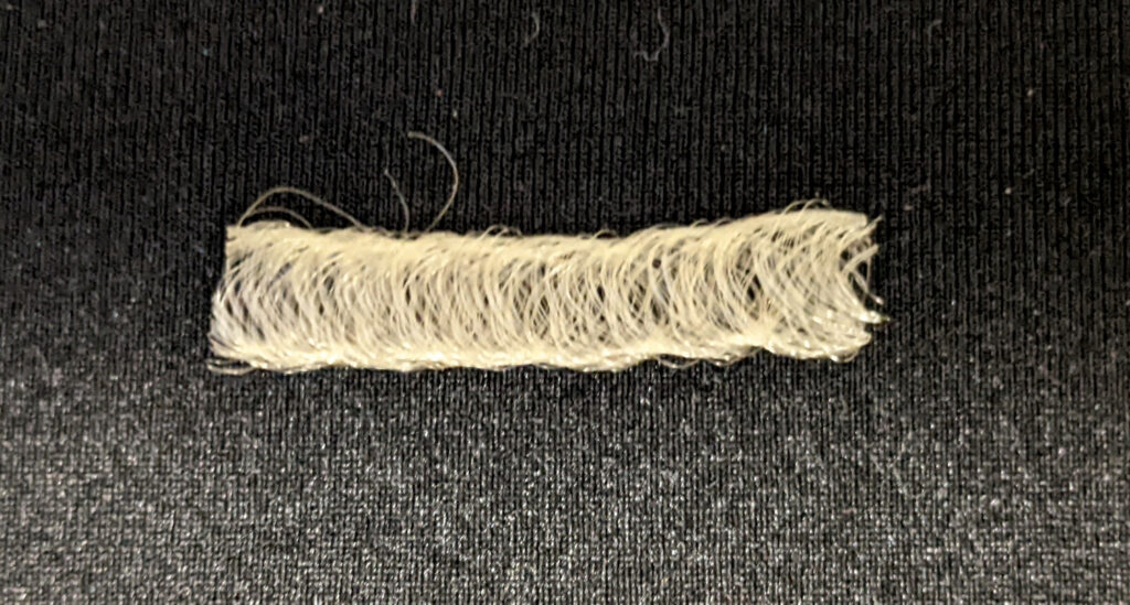
NCSU researchers are developing an implantable, resorbable mesh designed to be placed at the tumor site following surgery. The mesh is made from polylactic-co-glycolic acid (PLGA), a biodegradable and biocompatible polymer that is approved by the Food and Drug Administration (FDA) for drug delivery applications. The mesh is produced using a specialized braiding technique that results in a flexible, porous, tubular structure, providing mechanical support to the surgical site.

This structure is coated with a resorbable hydrogel containing doxorubicin. As the PLGA polymer degrades, it breaks down into lactic and glycolic acids—which are naturally metabolized by the body—avoiding the need for surgical removal. The hydrogel coating enables a sustained release of doxorubicin for two weeks, which is crucial during the postoperative window before scar tissue begins to hinder drug delivery. This localized delivery system effectively maintains therapeutic drug concentrations at the tumor site while minimizing systemic exposure and associated toxicity.
Studies have shown that more than 80% of the drug is released within the first two weeks, which coincides with the critical postoperative period. The mesh maintains its structural integrity for three weeks before gradually undergoing surface erosion, effectively balancing mechanical support with resorption timelines.
Initial in vivo studies in mice have demonstrated that the blank PLGA mesh and hydrogel are noninflammatory, biocompatible and safe for implantation. The researchers are working to improve the implant’s mechanical properties, drug-release kinetics and ease of surgical placement.
Upcoming research will assess the timing of cancer-cell death, DNA damage and the inhibition of cell proliferation. In vivo studies will focus on evaluating tumor reduction and recurrence rates. Early data indicate that this system could significantly decrease recurrence.
Collaborating with surgeons and industry partners is crucial to advancing this technology. Input from surgeons will enhance the design of the mesh to allow for customizable sizes and shapes, aid in the implantation process and validate its efficacy in preclinical trials. Obtaining regulatory approval will require conducting toxicity studies in larger animal test subjects and undertaking trials to establish safety for human use.
For successful commercialization, collaborations with biotechnology firms will accelerate clinical adoption. Furthermore, adapting this platform for other peritoneal cancers such as ovarian or gastric cancers will expand its market potential.
Composite scaffolds for skin injuries

The second project involves the development of 3D fiber-foam composite scaffolds for skin tissue engineering for treating skin conditions that have limited treatment options. These include skin injuries such as burns, chronic wounds, abdominal fistulas, diabetic ulcers and genetic defects.
When treatment fails, it can lead to infections and life-threatening complications. Autografts, which involve taking the patient’s own tissue, and synthetic skin substitutes have limitations such as donor site morbidity, poor integration and inadequate mechanical properties.
The research team’s approach is to develop 3D foam-fiber composite scaffolds that mimic the structural and mechanical properties of native skin tissue. The bioresorbable polymer foam matrix can be created to have a precisely controlled pore-size gradient through the thickness of the scaffold.
Fibers or yarns are added within the foam to adapt to various skin types, thicknesses and anatomical locations. The resulting 3D foam-fiber composites can then be surface-coated with collagen or gelatin to improve cell compatibility.
The work uses a repeatable and scalable manufacturing process. Individual parameters such as fiber type, pore-size distribution, overall dimensions and type of foam matrix can be selected and combined to build the desired foam-fiber composite structure. These structures have superior mechanical properties compared to unreinforced foams and provide excellent surfaces for cell migration, adhesion and proliferation.
These 3D foam-fiber composite scaffolds combine the advantages of different materials and structures. The fabrication method developed allows independent selection of the type of reinforcing fiber or yarn, the fiber or yarn density, and orientation, integrated within a polymer foam with a specific architecture. This approach allows the replication of the complex architecture of natural skin tissue, thereby improving the potential for successful skin tissue regeneration and function.
To develop this technology further and achieve commercial manufacturing production levels, it will be necessary to scale up fabrication. This will involve the development of improved tooling for the scaffold creation process and the polymer infusion and foam formation steps.
Animal testing will be necessary to determine the biocompatibility and durability as well as the selection of the optimal composite structure and degradation rate in an in vivo environment. This data will be required by the FDA for the Class 2 regulatory approval submission.
Barbed surgical sutures
The third project involves the development of a special type of barbed or knotless surgical suture used by plastic and cosmetic surgeons. The title of the project is “Ultrashort Pulse Laser Fabrication and Evaluation of Innovative Resorbable Barbed Sutures.”
According to the American Society for Plastic Surgery, there were 1 million reconstructive procedures performed in the U.S. in 2023, and this number is growing worldwide. To achieve better surgical outcomes, plastic surgeons are transitioning from a conventional knotted monofilament and braided surgical suture to a knotless suture known as a barbed suture. These barbed sutures have a plurality of barbs cut at specific intervals around the periphery of the main filament.
Currently, these barbed sutures are manufactured mechanically using a straight metal blade with the same standard barb dimensions and geometry. However, the same barb cut depth and cut angle are not suitable for all surgical procedures. The anchoring performance of a barbed suture varies depending on the surrounding tissue, the anatomical site and the procedure.
In an experimental study, barbed sutures were fabricated in various shapes and geometries using a femtosecond laser, which is an ultrashort pulse laser system. Both straight and curved barbs were fabricated on resorbable catgut and synthetic biomaterial monofilaments, such as poly-4-hydroxybutyrate.
The femtosecond laser system fabricated both straight and curved barbs consistently with similar mechanical properties and anchoring behavior to those barbed sutures cut with a blade. The laser-cut barbs were more reproducible and had improved precision compared to the mechanically cut barbs. An evaluation of the thermal behavior and degradation profile of the barbed sutures confirmed that the femtosecond laser system had a negligible effect on the integrity of the polymeric material.
This work shows a promising direction for the commercial scale-up of barbed suture manufacture.
Currently, the research team is in discussion with Dr. Adam Summers, a plastic surgeon in Baltimore, Md., who is considering licensing this technology as a component in a new surgical device.
Essential for success
The research and development process often advances by working in an interdisciplinary manner, bringing different technologies together for the first time. The two essential ingredients in converting a novel idea into a viable commercial clinical or health care product are:
- A clinician, surgeon or health care professional who sees merit in the invention to be an ambassador to promote its clinical use.
- A business or corporate entity to collaborate with to ensure that the scale-up and launch are feasible from technical, legal, commercial, promotional, sales and marketing points of view.
For each of the three examples here, researchers at the Wilson College of Textiles continue to seek and include both ingredients as R&D advances through prototyping, in vitro evaluation and in vivo animal trials.
The co-authors are, or have been, members of the Biomedical Textiles (BMT) Research Group at the Wilson College of Textiles, North Carolina State University (NCSU).
Martin King, Ph.D., is a professor of biotextiles and textile technology at Wilson College of Textiles. He is also a fellow of the Institute of Textile Science in Canada and an associate (chartered engineer) of the Textile Institute in the U.K.
Mengnan Dennis, Ph.D., is a postdoctoral research scholar working in the BMT Research Group with Martin King, Ph.D., on developing fiber-foam composite scaffolds for skin tissue engineering.
Karuna Nambi Gowri, Ph.D., is a postdoctoral research scholar working in the Department of Forest Biomaterials NCSU College of Natural Resources with Joel Pawlak, Ph.D., an associate professor at the university.
Ummay Nisha Jahan is a doctoral candidate working with Martin King, Ph.D., and Dr. Andrea Hayes-Dixon, a pediatric oncology surgeon at Howard University, to develop implantable devices for targeted chemotherapeutic drug delivery.